
Diagnostic Imaging
X-rays
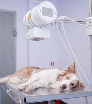
We at Ancu Vets use digital x-ray machines to quickly diagnose issues that might affect your pet..
A pet ultrasound is a non-invasive imaging technique used to view your pet's internal organs and identify potential issues.

Ultrasound
-
An ultrasound is a safe, non-invasive diagnostic procedure that gives your vet a two-dimensional ‘picture’ of your pet’s organs. In the procedure, a hand-held probe uses sound waves to create an image that’s displayed on a monitor.
-
It can be used to investigate issues such as:
a heart murmur
irregular heartbeats (arrhythmia)
laboured breathing
birth problems
organ diseases (e.g. liver and kidney problems)
vomiting and diarrhoea
foreign objects in the body
tendon injuries
pregnancy
urinary problems
-
It’s sometimes not necessary to anaesthetise or sedate your pet for ultrasound examinations as the technique is completely painless. Many pets will simply lie comfortably while the scan is being performed. If your pet is particularly sensitive or anxious, a sedative may be needed.
Your pet’s hair will generally need to be clipped over the area being examined to allow for a clear image. Your vet will then place gel on the area and methodically move the ultrasound probe around to record images of the area of interest.
Ultrasound is perfectly safe, and unlike an x-ray doesn’t expose your pet to any radiation.
FAQs Surrounding X-ray
-
An x-ray (or a radiograph) is a diagnostic procedure that lets us see inside your pet’s body to check their bones and organs.
It’s a common imaging technique that gives your vet more information about what’s going on inside your pet.
-
While x-rays can be used to identify fractured and broken bones after an injury, they can also diagnose many other issues.
They may be used to:
investigate orthopaedic issues such as lameness, fractures and deformities
assess a bloated or painful abdomen, vomiting or diarrhoea
view changes in your pet such as tumours, cysts or stones
check on conditions that affect the heart, lungs, liver and other organs
diagnose dental problems, such as tooth root abscesses and fractures.
-
The x-ray equipment will be placed over the part of your pet’s body to be examined in order to get the required images. Your pet will usually need to be re-positioned to show several different angles.
Your pet will need to be sedated or anaesthetised in order for an x-ray to be taken.
If you have any questions about x-rays at Medivet, contact your local practice.